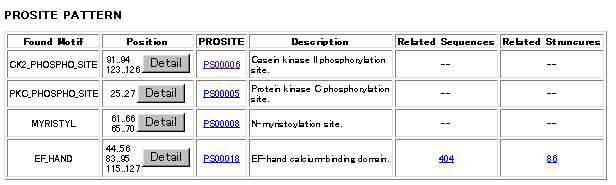
PROSITE is a database of protein families and domains. It consists of biologically significant sites, patterns and profiles that help to reliably identify to which known protein family (if any) a new sequence belongs.
This server allows you to search a single sequence againt PROSITE database using program MotifFinder written in ICR, Kyoto University. It not only finds out sequence motifs in your query sequence, also provides functional and genomic information of the found motifs using DBGET and LinkDB as the hyperlinked annotations. The results will also be presented graphically, and especially, wherevere available, 3D structures of the found motifs can be examined by RasMol program.
It first searches PROSITE pattern library of sequence motifs for patterns that appear in your query sequence. Common sequence patterns such as "C-kinase phosphorylation site" or " N-glycosylation site" (which are assigned as "/SKIP-FLAG=TRUE" on the CC line in the database) are ignored automatically.
If any patterns are found in the database MotifFinder then looks up the sequence entries annotated in PROSITE dabase, Swiss-Prot
entries which are described on DR lines ("True positive" ones only)
as well as PDB entries described on 3D lines.. Former entries are again searched
with the motif pattern found along with other motifs described on FT lines (Feature Table)
in the Swiss-Prot entries. Those results are shown as
graphical feature diagrams under Related Sequences column and can be seen by clicking the number of
sequences found shown in the table.
In the feature diagram a pink box shows the position of the motif pattern found, as well as red, yellow and green
lines represent active, binding and modification sites respectively.
| Colour | Sequence pattern |
| Pink | Motif Pattern |
| Red | Active site |
| Yellow | Binding site |
| Green | Modification site |
Under Related Structures column you can find the number of three dimensional structures available in Protein Data Bank which share structural region(s) with motif pattern found in PROSITE database. A list of those entries, ID numbers of PDB together with brief description of the proteins is shown by clicking the cell. From the list you can either jump into DBGET to look at the PDB entry more precisely, or see the position of the motif pattern on the 3D structure.
Under Position column of the table the position (start and end sequence numbers) of found motifs are list. Click Detail bottun to see actual positions of the motif patterns along the query sequence .
Below a sample output table of the search is shown. Click the images to get detailed results discribed above.
|
|